DECONV
Differential haemodynamic responses in the human striatum during initial and reversal learning
🧠 Contrary to common assumptions, BOLD fMRI does not measure neuronal activity directly. Rather, it measures a signal proportional to the ratio of oxy to deoxy haemoglobin. As such, we are actually instead measuring blood flow, a reliable proxy for neuronal activity as neurons require oxygen delivered through the blood in periods of higher activity.
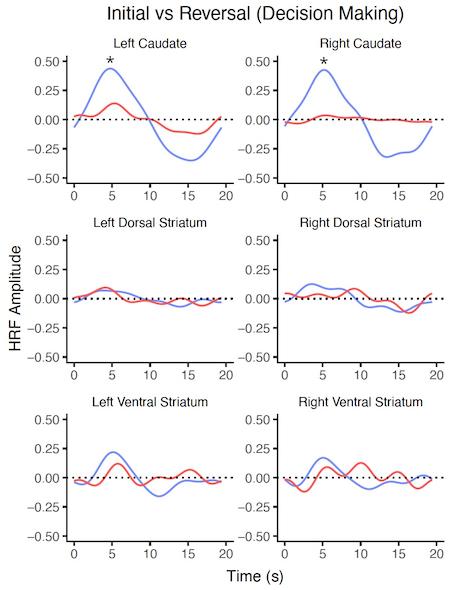
📈 Therefore, a vital component of modelling the BOLD response is the blood flow, commonly modelled using a haemodynamic response function (HRF). The HRF is therefore important when understanding the temporal dynamics of the neural response, as you gather information not inferrable simply from the BOLD statistics. Whilst BOLD maps will tell you what regions of the brain demonstrate more activity that others, deriving the HRF provides information regarding the intensity of this response (time-to-peak), the magnitude of the response (peak height), and the size of the response (full-width half-maximum).
Therefore, in this project we re-analysed data from a reversal learning task, performing deconvolution to extract HRFs from regions of the striatum, during feedback and decision making epochs in initial learning and reversal learning phases. We were able to demonstrate significant changes in the HRF between initial and reversal phases for both decision making and feedback epochs in the caudate, suggesting a functional role of this region with initial but not reversal learning.
Click here to see a full-screen version of a poster summarising the project presented at the Interpreting BOLD (Online) conference.
🌐 Team:
University of Reading:
Professor Anastasia Christakou (Centre for Integrative Neuroscience and Neurodynamics)
Dr Gabriella Rossetti (Centre for Integrative Neuroscience and Neurodynamics)
External:
Dr Tiffany Bell (University of Calgary)